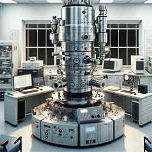
As my group does a lot of high resolution microscopy, we realized in a cross-sectional TEM work on silicon wafers had nanosized crystallites grown in 300 nm amorphous SiO2 layer. This could be a part of the problem why color looks different in different wafers.
How Scientists Use TEM Microscopy for Their Research
A university researcher used our substrates for their TEM work.
Buy Just One Thermal Oxide Silicon Wafers! Start Researchign Today!
Please reference #ONLQ25016 for quote/specs.
Get Your Quote FAST! Or, Buy Online and Start Researching Today!
What is the Best Applications for a Transmission Electron Microscope?
Transmission Electron Microscopy (TEM) is a powerful imaging technique with many applications across a variety of fields, including materials science, biology, physics, and chemistry. Some of the best applications for TEM include:
-
Material science: TEM is commonly used to study the microstructure and defects of materials, including metals, ceramics, polymers, and composites. TEM can provide detailed information on the arrangement of atoms, the presence of impurities and defects, and the crystalline structure of the material.
-
Nanotechnology: TEM is a critical tool for the study of nanoscale materials, such as nanoparticles, nanocomposites, and nanostructured surfaces. TEM allows for the imaging and characterization of the structural, chemical, and electrical properties of these materials at the nanoscale.
-
Biology: TEM is used in the field of biology to study the ultrastructure of cells and tissues, including the arrangement of organelles, the structure of proteins, and the behavior of viruses.
-
Materials characterization: TEM is used to characterize the properties of materials, including the electrical conductivity, magnetic properties, and chemical composition.
-
Electron diffraction: TEM can be used to study the crystal structure of materials by using electron diffraction patterns. This information is critical for understanding the properties and behavior of materials.
-
Environmental scanning electron microscopy: TEM is used in environmental scanning electron microscopy (ESEM) to study the surface structure and composition of materials in their native state, without the need for vacuum conditions.
In summary, TEM is a versatile and powerful tool with many applications across a variety of fields. Its ability to provide high-resolution imaging and detailed information on the structure of materials at the nanoscale makes it a valuable tool for a wide range of research and industrial applications.
How Does Transmission Electron Microscope Work with Silicon Substrates?
Transmission Electron Microscopy (TEM) is a powerful tool for imaging materials at the nanoscale. When used with silicon substrates, TEM works by shining a highly focused beam of electrons through a thin, transparent sample. As the electrons pass through the sample, they are either transmitted or scattered by the atoms in the material, forming an image of the internal structure.
To prepare a silicon substrate for TEM imaging, the material is typically thinned down to a few tens of nanometers or less. This can be done by various techniques such as mechanical or chemical polishing. The thinned sample is then mounted onto a support grid and placed in the TEM instrument.
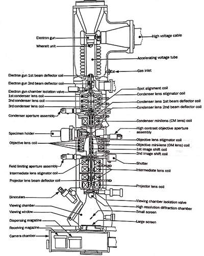
In the TEM instrument, a high-energy electron beam is generated and passed through a series of lenses that focus the beam down to a very small, tightly focused probe. This probe is then directed towards the sample, and the electrons that pass through the sample are detected by a specialized detector, such as a camera or a scintillator screen. The resulting image provides detailed information about the internal structure of the material, including the arrangement of atoms and any defects or impurities.
When using TEM with silicon substrates, it is important to consider that silicon is a relatively heavy atom and therefore scatters the electrons significantly. This can make it challenging to obtain high-resolution images, as the electrons are deflected by the silicon atoms and may not reach the detector. However, advances in TEM instrumentation and sample preparation techniques have made it possible to obtain high-resolution images of silicon and other materials, providing valuable insights into their structure and properties.
Magnification ofTransmission Electron Microscope
The magnification of a Transmission Electron Microscope (TEM) is determined by the combination of the electron optics and the imaging detectors used in the instrument. The electron optics, consisting of electromagnetic lenses and aperture systems, control the size and shape of the electron probe and determine the final magnification of the image. The imaging detectors, such as cameras or scintillator screens, collect and convert the transmitted electrons into a visual image.
TEMs can achieve extremely high magnifications, often on the order of millions or even billions of times. The maximum achievable magnification is limited by the quality of the electron optics and the size of the electron probe, which is directly proportional to the resolution of the image. The higher the resolution, the smaller the electron probe and the higher the magnification.
In practice, the actual magnification used in a TEM depends on the specific requirements of the sample being studied. High magnification is typically used to image small features or to resolve details within a material, while lower magnification is used to image larger structures or to survey the overall sample structure. The user can adjust the magnification by adjusting the electron optics and imaging detectors, or by selecting different operating modes in the instrument.
In summary, TEMs are capable of achieving extremely high magnifications, and the actual magnification used depends on the specific requirements of the sample being studied and the instrument's capabilities.
How Does a Transmission Electron Microscope Work?
Transmission electron microscopy microscopes use electron beams to create atomic-tem resolution images of objects. The main feature is its ability to detect the presence of atomically small particles and can study living things. .
Transmission electron microscopy
A transmission electron microscope (TEM) is a type of electron microscope that uses electrons to image biological samples. It is a high-resolution instrument that is ideal for a multiuser facility. The final magnification of a TEM image is usually 1,000 to 250,000 times the original magnification. Higher magnifications can be obtained through photographic or digital enlargement techniques. The final image quality depends on the accuracy of the lenses and the electronic stabilization system.
Transmission electron microscopy is useful for forming images of atomic arrangements in localized regions in a material. It can reveal the microstructure and interfaces of complex materials. The technology plays an important role in the development of new materials. Furthermore, it provides important information on the macroscopic properties controlled by defects and interfaces. In addition, transmission electron microscopy can be used to complement other techniques in materials science. In addition to transmission electron microscopy, X-ray and neutron diffraction are useful for determining the average structure of a complex material. In addition, they can reveal the local and individual nanostructures of a sample.
A transmission electron microscope uses several lenses in order to produce an image. The samples are located on a movable specimen stage. A short-focus lens is used to create the real intermediate image, while a projector lens magnifies the image. A single projector lens can provide a range of 5:1 magnification, but a modern microscope uses two projector lenses for better magnification.
It uses electron beams instead of light
The electron beam, which is negatively charged, interacts with the sample atoms and carries away compositional and contrast information. This information cannot be interpreted by human eyes, but special electron imaging devices can convert the electron signal into a greyscale image. These images correspond to the differences in the density and thickness of the sample being examined.
It uses diffraction to obtain atomic-resolution images
Transmission electron microscope (TEM) is a powerful tIn a TEM, electrons pass through the sample, scattering due to an electrostatic potential. The electrons then pass through an electromagnetic objective lens, which focuses the scattered electrons into a single point. This image is then projected onto a screen or camera.ool for studying specimens in atomic detail. Unlike conventional electron microscopes, TEM makes use of diffraction to obtain atomic-resolution images. These images are directly interpretable because they show the distribution of atomic species within a specimen. The resulting images also allow researchers to identify the specific chemical species present in a specimen.
Different TEM lenses work by focusing the beam from the sample. A single-lens system produces a parallel beam with a diameter greater than 1 millimeter, while a double-lens system produces a highly focused beam smaller than an atom.
Coherent diffraction imaging experiments can improve the accuracy of TEM images. With the use of electron sources with higher coherence, the resolution of atomic images can be improved. The diffraction data can be used to study interfaces between two materials, or defects in crystals. This type of imaging also requires well-controlled procedures to minimize lens aberrations.
It can be used to study living things
A Transmission Electron Microscope beam allows for greater magnification and resolution biological objects. However, it is more expensive and bulky than a light microscope. The Transmission Electron Microscope can be used to study living things and is an essential tool for scientists.
The transmission electron microscope is a device used in science and medicine to study the structure and composition of living things. It uses a focused beam of electrons, usually five to 100 keV, to investigate a specimen. The beam is then processed and converted into an image.
It uses a holy grid
A holey grid is an imaging enhancement technique that optimizes the imaging capabilities of a Transmission Electron Microscope. This imaging aid consists of a TEM grid support that is covered with a thin plastic film or stabilizing carbon layer. It contains tiny round holes that create Fresnel fringes when the electron beam passes through it at high focus. The banding effect is the result of electrons diffracting around the edges of the holes.
The amorphous film withstands the high vacuum inside the electron microscope. This property helps to preserve the native structure of the sample, which is crucial for high resolution imaging. Because the film is thin, it must be maintained at temperatures below -150 degrees Celsius to prevent devitrification. The objective aperture also enhances contrast.
Another important consideration when using the TEM is the sample's condition before being scanned. A thin sample layer is necessary to avoid extensive noise caused by scattered electrons. It should also be thick enough to withstand the high vacuum of the electron microscope. This thin layer will also protect the biomolecules from radiation damage. In addition, negative stain techniques adsorb the sample to a thin carbon film or embed it in an amorphous heavy metal. Once this has been completed, the assembly is allowed to dry in air.
It uses converging lenses
A Transmission Electron Microscope (TEM) uses converging lenses to focus light onto a sample specimen. The lenses are different from those used in an optical microscope, and are used to focus light onto an electron beam rather than a light source. The TEM can achieve extremely high magnifications. The resolving power of a TEM depends on the electron voltage. High electron voltages can achieve wavelengths as short as 5 nanometers and as long as 2 nanometers. These characteristics enable high magnifications, and the TEM has widespread applications in micrography of biological cells and viruses.
It uses amplitude contrast
The Transmission Electron Microscope is a popular method of studying biological samples. Its high resolution makes it useful for studying viruses, organelles, and macromolecules. The instrument requires a sample to be placed inside it, and the electron beam is then emitted. It can also be used to study special materials. This type of microscope is often used in conjunction with the light microscope.
A TEM image is not simple to read and understand at high magnification, and the spectral resolution is only one component of the overall contrast. The contrast transfer function of a TEM depends on the imaging parameters, and is a function of spatial frequency. The more defocus the specimen is, the lower the point resolution. However, it can reveal general morphology of the sample.
The wavelength of electrons that are used for transmission are very small. The wavelength is proportional to the accelerating voltage, and so the wavelength of an electron is shorter the higher the accelerating voltage. By applying these two equations to the TEM, the resolution can be calculated. Resolution is the distance at which two objects can be distinguished.
