We are looking for a source of high-purity silicon Wafers or ingots with p-type doping. The p-type doping should be very low, below 2E16 1/cm3 I noticed that in your catalog, there are only high-purity n-type Si. Please check if you can get high-purity, lightly boron-doped silicon wafers or ingots with low dislocation density. We are looking for a large quantity. Diameter 2-4.
What is Dislocation Density?
Silicon Wafers and Iingots With Low Dislocation Density
A Ph.D in Ph.D. electrical engineering requested help with the following:
Reference #285127 for specs and pricing.
Get Your Quote FAST! Or, Buy Online and Start Researching Today!
Dislocation Density of AlN on Sapphire Substrates
A posdoc requested help with the following:
I request a quote for: - un-doped AlN on Sapphire, c-axis orientated 2'' thikness 1000nm grown by MOCVD ( also price for m-axis orientated) - un-doped AlN on Sapphire, c-axis orientated 2'' thikness 2000nm grown by MOCVD. and i ask specifications details for each material essentially band gap, residual doping, dislocation density, resistivity.
Reference #262030 for specs and pricing.
Advantages and Disadvantages of Silicon Wafer Dislocation Density
The silicon wafer dislocation density is a crucial parameter for semiconductor manufacturing. It  is dependent on several factors, including the Ori of the silicon wafer. The most popular Ori is the vertical, since it is easier to cleve. However, there are many alternatives. Here are some advantages and disadvantages of each. Listed below are some other advantages and disadvantages of silicon wafers. This article will discuss the most common and practical approaches to evaluating wafer dislocation density.
Ori of silicon wafers influences the semiconductor properties. The optimum Ori depends on the specific requirements. Depending on the type of device, a semiconductor may be oriented horizontally, vertically, or in a non-uniform fashion. The differences between vertical and horizontal oriented silicon wafers are discussed. The Miller index is useful for determining the optimum Ori. The three most common types of silicon wafers are crystalline, amorphous, or epitaxial.
To determine silicon wafer dislocation density, use an opticmicroscope. A well-designed optical microscope can measure the density of dislocations. A 150 mm Czochralski grown wafer, for example, had a high boron doping level. A cell piece detecting instrument by the German halm company can also measure the photoelectric transformation efficiency. Its efficiency was 17.3%. A study of a thin film solar cell has shown that the p/p+ silicon wafers are more susceptible to misfits than the monolithic wafers.
Dislocation Density Video Tutorial
What is Dislocation Density?
Since dislocations are responsible for most plastic deformation phenomena in metals, it is important to know how they behave and the density of their clusters. Several instrumental techniques have been developed to measure dislocation density. X-ray diffraction and transmission electron microscopy (TEM) are two of the oldest methods to measure dislocation density. These two methods can be used to determine the number of dislocations and the number of clusters in the sample. However, these methods are not practical for large-scale investigations, as they require a large observation area and the samples must be extremely thin to count them.
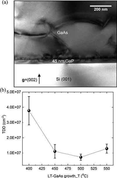
XRD and TEM are two of the most common methods for measuring dislocation density. A Weak-beam Dark-field image can be used to estimate dislocation density in a small cubic volume. This method is accurate in measuring localized density of dislocations and can provide a rough estimate of dislocation densities. The figure below shows the measurement results for XRD and TEM. It can also be used to calculate the thickness of the sample.
The PVScan tool measures the etching pit density, which is an index of dislocations per unit volume. The EPD is typically measured in the range of 10-4 to 3 x 10⁻⁶ cm-2, but lower or higher values do not show a detectable change. In small areas, where the dislocation density is low, the etching pit density will be low or will not be detected at all. If the cluster is large enough, the etching pit density may be very high.
In addition to using XRD and TEM to measure dislocation density, the TEM also has an advantage in imaging the periphery of the wafer. By analyzing the TEM image, the resulting density of misfit dislocations is calculated. This is particularly helpful when determining the density of dislocations in a small sample. In XRD and TEM, roughness is a key parameter in measuring the density of dislocations in crystals.
A typical graph of dislocations is similar to that of a crystal in a solid, with higher density showing fewer dislocations. It is important to note that a high dislocation density does not necessarily mean a weaker crystal. Despite its low dislocation density does not necessarily indicate a weaker crystal. Instead, it simply means that dislocations are more widespread in the material. This explains why a thin film with a higher dislocation density will perform better in a larger device.
The size of the individual dislocations is an important determinant of their density. The larger the sample, the more dislocations there are, and the more crystalline defects are. This process also makes the dislocation density of crystals appear larger. The data is used to determine how a material should be manufactured. If the process can be optimized, it can dramatically reduce the size of the process. This is a crucial step in making the semiconductor industry, as it improves the reliability and performance of semiconductors.
In order to determine the dislocation density of annealed samples, we used the Ham intercept method. This is an intercept counting method that takes into account the magnification of the images. We acquired four micrographs of the B85 and BA samples to determine their dislocation density. The values of the sample thickness were obtained by the Electron Energy Loss Spectroscopy. Then, we calculated the total area of the individual micrographs, and compared them.
The density of dislocations is also related to the roughness of the edges. While a surface is smooth and unbleached, roughness can cause a lot of dislocations. Thus, a smooth edge is necessary for the dislocation to propagate. A rough edge will lead to more misfits. A sharp edge will result in a smooth surface. This means that the edge of a wafer is not as rough as the other edges of the wafer.
The dislocation density of a material can be measured using a variety of techniques. XRD and TEM are the two most common tools for measuring the density of dislocations. Among these, a Weak-beam Dark-field image can be used to estimate the local dislocation density. The same imaging technique can be used to estimate the density of multiple crystals. Once the topography is taken, a graph of the data can be viewed.
