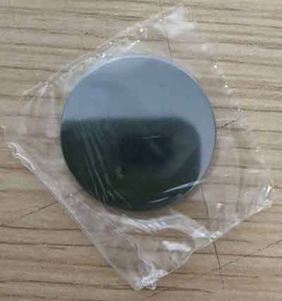
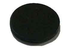
We would like to purchase several Si-specimen holders for XRD measurements. Our specifications are: 3,2 mm diameter ca. 2 mm thickness top side polished zero-background orientation without metal holder without cavity We would like to receive a quotation from you for 6 specimen holders.
X-ray Diffraction @ Zero Background Specimen Holder
A researcher from a university physics department requested a quote for the following:
UniversityWafer, Inc. Quoted:
Silicon single wafer single side polished
Diameter 32 mm
Thickness 2 mm
Central hollow: diameter 10 mm deep 0.2 mm
Application : X-ray diffraction @ zero background specimen holder
qty. 6 pcs
Reference #266431 for pricing.
Get Your Quote FAST! Or, Buy Online and Start Researching Today!
XRD Specimen Holders
Researcher:
I would like to purchase several zero-background XRD specimen holders compatible with Rigaku Miniflex XRD. Please show me some options and pricings.
would like a zero-background XRD sample holder for our  Rigaku Miniflex.
We are using ASC-8 automated sampler. I think if your holders are compatible, we would be happy to purchase from you.
Our Aluminum sample holder that is compatible with the ASC-8 sampler measures:
Inner dish diameter: 15/16"
Outer diameter: 1 9/32"
the holders are magnetic and stick well to ASC-8
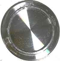
We would like to purchase 8 of these holders. I'm attaching two pictures just in case you are interested to see how our holder looks like.
I'm looking to see an XRD holder that does not show a signal over 20 - 90 degrees (2-theta) range.
Please let me know pricings for silicon and other options, if available. I'm looking to buy around 5 to 8 depending on the prices.
UniversityWafer, Inc. Provided The Following:
Item Qty. Description
HD38c. 5/10 Zero Diffraction Plate for XRD with pocket
Material: CZ Silicon,
Diameter: 32.0±0.3 mm,
Thickness: 2000±50 µm,
Flats: none, Edge rounding: straight,
Central pocket:
Diameter: 24.0±0.1 mm
Depth: ~500 µm,
Type/Dopant: p/Boron,
Resistivity: >10 Ohm-cm,
CTQ: Adequately oriented to guarantee zero background,
Surfaces: etched,
Adequately packed.
Contact us for pricing.
Researchers and X-Ray Diffraction
When we do X-ray diffraction. The little disc is just a platter that holds a small amount of powder in the divot. The platter is held in a larger metal or plastic ring that is the "sample holder". Most of the time, we use plastic for the platter, but the plastic gives off a small signal in the background. By using these Si wafers cut on specific crystallographic orientations, they are "forbidden symmetries", that is, they don't give any diffraction signal. Then we clean off the powder and do it again with the next material.
The following spec works.
Si single wafer single side polished
Diameter 32 mm
Thickness 2 mm
Central hollow: diameter 10 mm deep 0.2 mm
Application : X-ray diffraction @ zero background specimen holder
Please either send us your specs or let us know what quantity you can use.
What is X-Ray Diffraction Zero Background Sample Holder?
When you're diving into X-ray diffraction, a Zero Background Sample Holder is key—it lets you get crystal-clear data without any interference from the holder itself. X-ray diffraction, you know, it's like a detective's magnifying glass for scientists who want to get the lowdown on what stuff is made of at a molecular level. When we shoot X-rays at a material, the scattered rays tell us all about its internal structure.
A Zero Background Sample Holder is designed to minimize background noise in XRD measurements. In XRD,  background noise can come from various sources, including the sample holder itself. This background noise can muddle the XRD results, making it tough to pinpoint what's actually going on in the sample.
The Zero Background Sample Holder typically uses a single crystal, such as quartz or silicon, which has been cut in a specific orientation. Thanks to the single crystal's clean cut, it won't throw any extra peaks into our diffraction pattern analysis, keeping our results sharp and on point. As a result, it provides a 'zero background' or very low background, enabling more precise and accurate measurement of the diffraction patterns of the sample under investigation.
These holders are particularly useful for analyzing samples with very weak diffraction signals or for samples that are small or thin. By cutting down on the noise, they let us get sharper data that's key for nailing down how a material's crystals are put together and what makes them tick.
32mm x 2 mm holder XRD Sample Holder
An assistant professor of engineering requested a quote for the following:
Could you send me a quote for X-Ray Diffraction Zero Background Sample Holder?
Reference #278955 for specs and pricing.
What Is X-Ray Diffraction Zero Background Specimen Holder
There are several important tools to characterize the solid state character of drugs and  substances. In this section, we take a look at one of the most important of these tools: the X-ray Diffraction Zero Background Specification. [Sources: 6, 9]
Even if there is no crystallite size, the powder diffraction pattern can be calculated by simulating PXRD in micro- and nanocrystalline materials. Although a powder XRD is available, there is no information about the crystalline structure (or its absence). [Sources: 4, 9]
A simplified variant of approach 2 is to use the peak intensity of a single XRD peak at two theta positions where the amorphous signal is present but there are no overlapping crystalline X-RD peaks. It should be the base intensity (the maximum intensity), which lacks a significant signal (ammorphic), which can be used as the starting line for the subtraction of the isomorphic signal by the peak intensity of the crystalline of the XRAD. [Sources: 9]
In this way, we can determine the proportion of amorphous portions, and samples produced using this method will certainly be sufficient. Rietveld based method, which uses the entire diffraction pattern, can also be used to estimate the total volume of XRAD (or the amount of crystalline X-RD) in the sample if one is available. [Sources: 2, 9]
The sample holder rotates in its own plane, presents itself perpendicular to the X-ray beam and rotates around itself. The silent object then becomes a powder holder in which the shape of the sample is well defined - provided it is on the same plane as the specimen. [Sources: 7, 8]
The deviating slits decrease when the scan passes through higher diffraction angles, resulting in different bent angles. At lower angles, the sample intercepts only a portion of the beam, resulting in a decreased beam intensity and increasing the probability that the background will be scattered from the sample holder. Finally, there can be no effective suppression of example transparency (see next section). This results in an error in the estimation of the amorphous process, but at low angles it can also intercept only parts of the beam, resulting in a reduced X-ray intensity and thus a higher detection probability. [Sources: 5, 9]
This sensitive error can also be introduced into the X-ray beam by preparing the sample for presentation, for example by using a high-speed camera or laser beam. [Sources: 5]
One option is to glue or stick a microscope objective on the back of the sample holder. Bruker has a product sheet entitled "X-ray diffraction without background sample holder" which contains information about the X-ray background sample holder and its use. The AXRD worktop can hold a sample holder, but sample holders can also be improved by using standard Hull, Debye or Scherrer powder analysis. [Sources: 3, 4, 7]
Some instrument manufacturers offer their own sample holders, which consist of circular wells that are processed for different sample quantities. Other discs, known as Zero Background Holders (ZBHs), are made of plastic, creating the Bragg Peak Reflection Mode. [Sources: 8]
The typical diffraction range is within the routine profile measurement PXRD, but depending on the diffraction ometry and optics, the ZBH has a wide range of possible orientations to avoid background noise, which is evenly distributed in all possible orientations. Since electron crystallography requires that diffraction data be collected even from highly inclined crystals (70 degrees), where the effective thickness of the crystal is twice as high as the actual thickness, this further increases the background intensity of the noise. As a result, the diffraction pattern will appear in a different direction than the normal diffraction pattern due to the thickness difference. [Sources: 1, 6, 9]
A check of the blank scan would make it possible to estimate the instrumental background and subtract it from the scan. In addition, analysts should discuss the history of a sample with its submitter to understand whether the determination on amorphous content is appropriate. [Sources: 9]
Plates - such as crystals - lie flat in a sample holder, for example, but very few have a vertical orientation. Note: It is often better to use a background plate without background for Si and SiO2 single crystals as sample holder. If the sample is prepared with Azero background plates, a blank scan would be used instead. Some common zerobackground plates are: X-ray, X-ray and X-ray diffraction zeroplates. [Sources: 0, 4, 5, 9]
This is essentially required to keep the sample on a tangent to the focus circle, but there is no other object provided the holder does not obstruct the X-rays and does not scatter them as they hit the powder sample. [Sources: 5, 7]
Sources:
[0]: http://science.sjp.ac.lk/instrumentcenter/instruments/rigaku-ultima-iv-x-ray-diffractometer/
[1]: https://www.cell.com/biophysj/references/S0006-3495(02)75619-1
[2]: http://www.ccp14.ac.uk/solution/powdermounts.htm
[4]: http://www.ilpi.com/inorganic/glassware/xrd.html
[5]: http://clay.uga.edu/courses/8550/XRD.html
[7]: https://patents.google.com/patent/US2317329A/en
[8]: https://onlinelibrary.wiley.com/iucr/itc/Ha/ch7o2v0001/sec7o2o3o2/
[9]: http://www.americanpharmaceuticalreview.com/Featured-Articles/36758-Approaches-to-Quantification-of-Amorphous-Content-in-Crystalline-Drug-Substance-by-Powder-X-ray-Diffraction/
